For an optimal
daily experience
daily experience
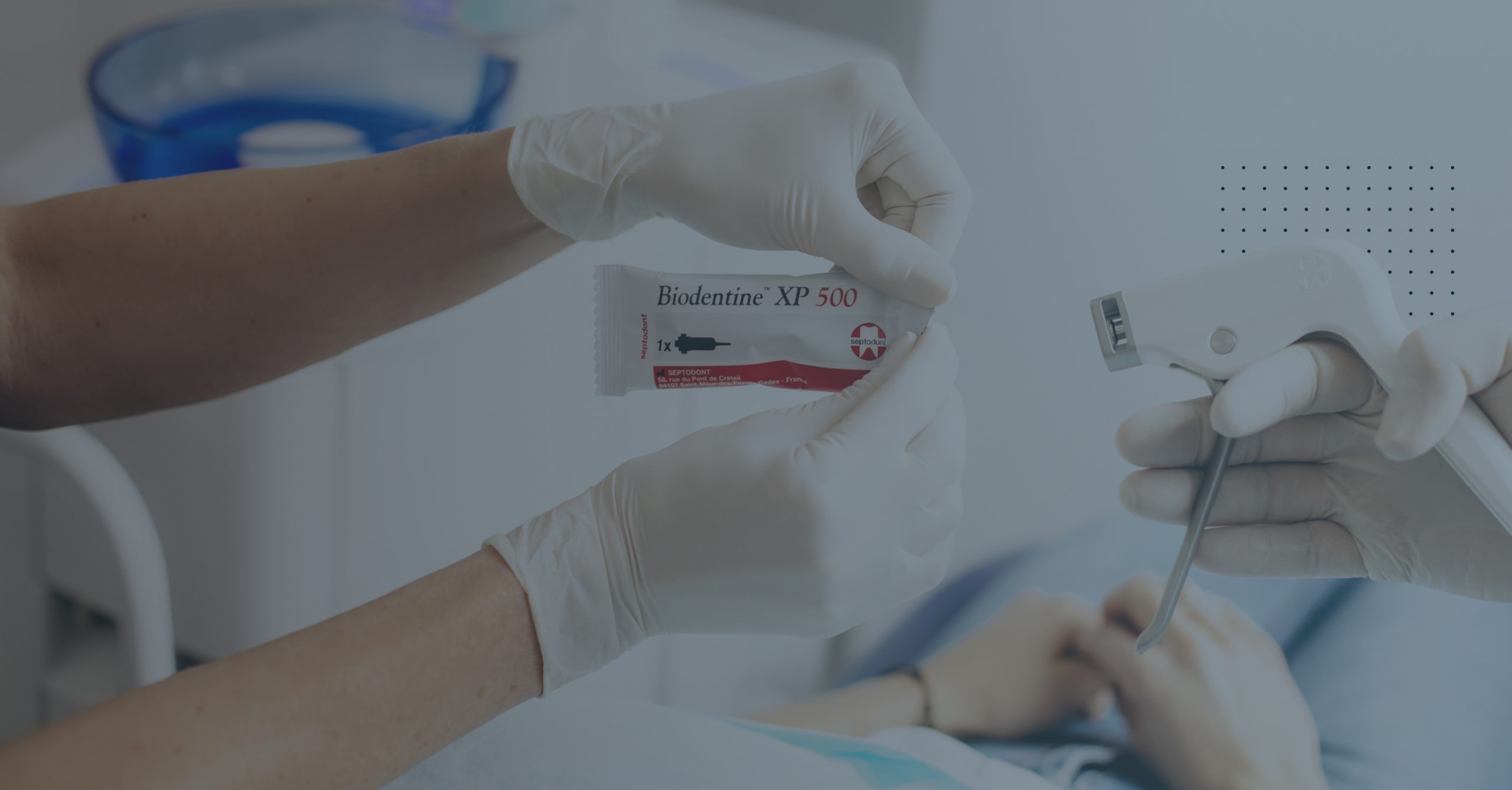
The ease of use and multiple indications of Biodentine from the crown to the root will help you every day to treat your patients
SEE ALL INDICATIONS
To learn more about Biodentine
SEE ALL INSIGHTS
-
Clinical case 05 / 04 / 2023 Case Studies Collection 1
Discover clinical cases reported by dental professionals around the world who share their experience and the benefits of using Septodont innovations in their daily practice. View Case Studies PDF - 3.4 MO
-
Clinical case 31 / 03 / 2023 Case Studies Collection 19
Discover clinical cases reported by dental professionals around the world who share their experience and the benefits of using Septodont innovations in their daily practice. View Case Studies PDF - 3.6 MO
-
Clinical case 06 / 06 / 2022 Case Studies Collection 20
Discover clinical cases reported by dental professionals around the world who share their experience and the benefits of using Septodont innovations in their daily practice. View Case Studies PDF - 0.5 MO
1 Immediate enamel restoration
FIRST SESSION
Assess pulp vitality by the usual tests.
- Isolate the tooth with a rubber dam.
- Remove the infected dentine with a round bur and/or a hand excavator. Leave the affected dentine.
- Adapt a matrix around the tooth if a wall is missing.
- After activating the Biodentine XP cartridge, use Biodentine Mixer to ensure a perfect mix (refer to the instructions of use).
- Dispense Biodentine XP in the cavity using Biodentine Gun (refer to the instructions of use), so that the volume of missing dentine is replaced by the same volume of Biodentine XP avoiding trapping air bubbles. Flatten the material without excessive pressure and ensure good adaptation to the cavity walls and margins.
- Wait until the end of the setting time before performing the permanent enamel restoration.
Biodentine XP is compatible with all direct crown restoration techniques and particularly with all types of bonding systems.
2 Non-immediate enamel restoration
FIRST SESSION
Assess pulp vitality by the usual tests.
- Isolate the tooth with a rubber dam.
- Remove the infected dentine with a round bur and/or a hand excavator. Leave the affected dentin.
- Adapt a matrix around the tooth if a wall is missing.
- After activating the Biodentine XP cartridge, use Biodentine Mixer to ensure a perfect mix (refer to the instructions of use).
- Dispense Biodentine XP in the cavity using Biodentine Gun (refer to the instructions of use), avoiding trapping air bubbles. Ensure good adaptation of the material to the cavity walls and margins. Do not apply excessive pressure on the material.
- Model the surface of the restoration.
- Wait until the end of the setting time before removing the matrix.
- To optimise the mechanical properties of the material and facilitate removal of the matrix, a varnish can be applied onto the surface of the restoration.
- Check occlusion.
SECOND SESSION (1 week to 6 months later)
Within one week to six months after placement of Biodentine XP, prepare the cavity according to the criteria recommended for the selected restorative material.
The remaining Biodentine XP material can be considered as sound artificial dentine and permanently left in deep areas of the cavity and in areas adjacent to the pulp chamber. Biodentine XP is compatible with all direct or indirect crown restoration techniques (Inlay/Onlay), and particularly with all types of bonding systems.
3 Pulp capping (direct and indirect)
FIRST SESSION
Assess pulp vitality by the usual tests.
- Isolate the tooth with a rubber dam.
- Remove the infected dentine with a round bur and/or a hand excavator.
- Adapt a matrix around the tooth if a wall is missing.
- If there is bleeding in the pulp, haemostasis must be achieved before applying Biodentine XP.
- After activating the Biodentine XP cartridge, use Biodentine Mixer to ensure a perfect mix (refer to the instructions of use).
- Dispense Biodentine XP in the cavity using Biodentine Gun (refer to the instructions of use), so that the volume of missing dentine is replaced by the same volume of Biodentine XP avoiding trapping air bubbles. Ensure good adaptation of the material to the cavity walls and margins. Do not apply excessive pressure on the material.
- Perform the immediate or non-immediate enamel restoration as indicated above.
In case of non-immediate enamel restoration, a second session will be required.
Patients should be followed according to current recommendations.
4 Pulpotomy (reversible & irreversible pulpitis)
FIRST SESSION
Assess pulp vitality by the usual tests. In case of clinical signs and symptoms of irreversible pulpitis, pulpotomy is recommended when bleeding can be controlled in 5 minutes.
- Isolate the tooth with a rubber dam.
- Remove the infected dentine with a round bur and/or a hand excavator.
- Gain access to the pulp chamber and clean out the pulp. Gain access to the pulp chamber and clean out the pulp.
- If there is bleeding in the pulp, haemostasis must be achieved before applying Biodentine XP. If haemostasis cannot be achieved after 5 minutes, further pulp tissue should be removed (partial or full
pulpotomy) step by step until a controlled bleeding. A full coronal pulpotomy can be carried out to the level of the root canal orifices with bleeding arrested. - Adapt a matrix around the tooth if a wall is missing.
- After activating the Biodentine XP cartridge, use Biodentine Mixer to ensure a perfect mix (refer to the instructions of use).
- Dispense Biodentine XP directly in the pulp chamber using Biodentine Gun (refer to the instructions of use) and ensure good adaptation to the cavity walls and margins.
- Model the surface of the restoration.
- Wait until the end of the setting time before removing the matrix.
- To optimise the mechanical properties of the material and facilitate removal of the matrix, a varnish can be applied onto the surface of the restoration.
- Check occlusion.
SECOND SESSION (1 week to 6 months later)
- Within one week to six months after placement of Biodentine XP, prepare the cavity according to the criteria recommended for the selected restorative material.
- Patients should be followed according to current recommendations. The remaining Biodentine XP
material can be considered as sound artificial dentine and permanently left in deep areas of the cavity and in areas adjacent to the pulp chamber. Biodentine XP is compatible with all direct or indirect crown restoration techniques, and particularly with all types of bonding systems.
5 Repair of root perforations
FIRST SESSION
- Isolate the tooth with a rubber dam.
- Prepare the root canal alternately using suitable endodontic instruments and a solution of sodium hypochlorite.
- Dry the canal with paper points without totally dehydrating the root canal and use a calcium hydroxide
paste for disinfection between visits. Tightly seal the access cavity with a temporary cement to protect the temporary filling.
SECOND SESSION (1 week later)
- At the next visit (usually after one week) and if no symptoms, place a rubber dam and remove the temporary crown restoration. Clean the canal alternately using a solution of sodium hypochlorite and suitable endodontic instruments. Dry the canal with paper points without totally dehydrating the root canal.
- After activating the Biodentine XP cartridge, use Biodentine Mixer to ensure a perfect mix (refer to the instructions of use).
- Extrude Biodentine XP with Biodentine Gun (refer to the instructions of use) on a dental pad. Then, insert Biodentine XP in the perforation area using a suitable instrument.
- Condense Biodentine XP with a plugger.
- Take an X-ray to check that the material is correctly positioned.
- Remove excess material and place a temporary filling.
THIRD SESSION
Complete root canal treatment at the next visit according to current recommendations.
6 Repair of furcation perforations
FIRST SESSION
- Isolate the tooth with a rubber dam.
- Rinse the cavity with a solution of sodium hypochlorite to disinfect the area.
- If there is bleeding, haemostasis must be achieved before applying Biodentine XP.
- Dry the pulp chamber.
- After activating the Biodentine XP cartridge, use Biodentine Mixer to ensure a perfect mix (refer to the instructions of use).
- Depending on the size of the cavity, you can use Biodentine Gun either to dispense Biodentine XP
directly in the cavity, or to extrude the product on a dental pad and then apply it with a suitable instrument. Perforation repair and crown restoration are performed in a single step. - Take an X-ray to check that the material is correctly positioned.
- Remove excess material.
SECOND SESSION
At a subsequent visit, if all clinical signs of a successful treatment are present, the possibility of a permanent restoration can be considered.
7 Repair of perforating internal resorptions
FIRST SESSION
- Isolate the tooth with a rubber dam.
- Prepare the root canal alternately using suitable endodontic instruments and a solution of sodium hypochlorite.
- Dry the canal with paper points without totally dehydrating the root canal and use a calcium hydroxide
paste for disinfection between visits. Tightly seal the access cavity with a temporary cement to protect the temporary filling.
SECOND SESSION (1 week later)
- At the next visit (usually after one week) and if no symptoms, place a rubber dam and remove the temporary crown restoration. Clean the canal alternately using a solution of sodium hypochlorite and suitable endodontic instruments. Dry the canal with paper points without totally dehydrating the root canal.
- After activating the Biodentine XP cartridge, use Biodentine Mixer to ensure a perfect mix (refer to the instructions of use).
- Extrude Biodentine XP with Biodentine Gun (refer to the instructions of use) on a dental pad. Then, insert Biodentine XP in the perforation area using a suitable instrument.
- Condense Biodentine XP with a plugger.
- Take an X-ray to check that the material is correctly positioned.
- Remove excess material and place a temporary filling.
THIRD SESSION
Complete root canal treatment at the next visit according to current recommendations.
8 Apexification
FIRST SESSION
- Isolate the tooth with a rubber dam.
- Prepare the root canal alternately using suitable endodontic instruments and a solution of sodium hypochlorite.
- Dry the canal with paper points without totally dehydrating the root canal and use a calcium hydroxide
paste for disinfection between visits. Tightly seal the access cavity with a temporary cement to protect the temporary filling.
SECOND SESSION (1 week later)
- At the next visit (usually after one week) and if no symptoms, place a rubber dam and remove the temporary crown restoration. Clean the canal alternately using a solution of sodium hypochlorite and suitable endodontic instruments. Dry the canal with paper points without totally dehydrating the root canal.
- After activating the Biodentine XP cartridge, use Biodentine Mixer to ensure a perfect mix (refer to the instructions of use).
- Extrude Biodentine XP with Biodentine Gun (refer to the instructions of use) on a dental pad. Then, insert Biodentine XP in the apical area using a suitable instrument.
- Condense Biodentine XP with a plugger.
- Take an X-ray to check that the material is correctly positioned.
- Remove excess material and place a temporary filling.
THIRD SESSION
Complete root canal treatment at the next visit according to current recommendations.
9 Root-end filling in endodontic surgery
- Following apical resection, gain access to the operative site following the current recommendations in endodontic surgery.
- Using a specific ultrasonic tip, prepare a root-end cavity, 3 to 5 mm deep in the apical portion of the root canal.
- Isolate the area. Achieve haemostasis. Dry the cavity with paper points.
- After activating the Biodentine XP cartridge, use Biodentine Mixer to ensure a perfect mix (refer to the instructions of use).
- Extrude Biodentine XP with Biodentine Gun (refer to the instructions for use) on a dental pad. Then, insert Biodentine XP in the cavity in the root extremity using a suitable instrument. Condense Biodentine XP with a small plugger.
- Remove excess material and clean the surface of the root.
- Take an X-ray to check that the material is correctly positioned then close the area.
10 Revitalisation
FIRST SESSION
Assess pulp vitality and apex diameter by the usual tests. Revitalisation procedure is indicated for an immature tooth with necrotic pulp.
- Isolate the tooth with a rubber dam.
- Remove loose or necrotic pulp tissue using suitable endodontic instruments. Avoid mechanical instrumentation of the root canal walls.
- Irrigate with sodium hypochlorite solution (advised concentration: 1.3 to 3%, 20 mL, 5 min), use of side-vented needle, place 2 mm above vital tissue.
- Irrigate with sterile physiological saline (5 mL) and dry with paper points. Dry then with EDTA (advised concentration: 15 – 17%, 20 mL).
- Insert a non-discoloring calcium hydroxide product homogenously into the root canal. Instead of calcium hydroxide, a triple antibiotic paste consisting of ciprofloxacin, metronidazole and minocycline can be used.
- Place coronal seal directly onto canal dressing with a minimum thickness.
SECOND SESSION (usually 2 to 4 weeks later)
- Anaesthesia with a local anaesthetic without vasoconstrictor. Remove the temporary seal. Irrigate with EDTA (advised concentration: 15 – 17%), use a side-vented needle, place 2 mm above vital tissue. Irrigate with sterile physiological saline (5 ml). Remove excess liquid with paper points
- Induce mechanical bleeding of periapical tissue and rotational movement of an apically pre-bent file. Allow the canal to fill blood until 2 mm below the gingival margin, wait for blood clot formation for approximately 15 minutes.
- After activating the Biodentine XP cartridge, use Biodentine™ Mixer to ensure a perfect mix (refer to the instructions of use).
- Cut a collagen matrix to a diameter larger than the coronal part of the root canal and a height of 2 – 3 mm, place on top of the blood clot, allow the matrix to soak with liquid, avoid formation of a hollow space.
- Dispense Biodentine XP directly directly on top of the collagen matrix using Biodentine™ Gun (refer to the instructions of use) in a thin homogenous layer of about 2 mm underneath the cement-enamel junction.
- Then fill the cavity with Biodentine™ XP.
THIRD SESSION
On a next visit, refresh the cavity walls with a diamond bur or grit blast with aluminum oxide and seal. Complete with final restoration.